A first-of-its-kind instrument at Brookhaven National Laboratory (Upton, N.Y.) combines four imaging techniques into one experimental setup, enabling comprehensive studies of materials for next-generation nuclear reactors.
Mehmet Topsakal, a materials scientist in the Nuclear Science and Security Department at the U.S. Department of Energy’s (DOE) Brookhaven National Laboratory, and his team are leveraging high-energy x-rays to unveil different characteristics of nuclear materials, like their internal structure and how their elements are distributed after exposure to extreme environments. One class of x-ray techniques—computed tomography (CT)—builds a 3D picture of a material’s internal structure without destroying it or cutting it open. But scientists often need to conduct several different CT experiments to build a comprehensive understanding of their sample.
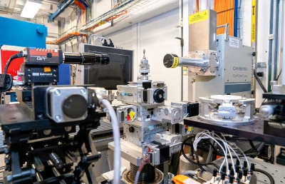
That’s why a team of Brookhaven Lab researchers has developed and built a single experimental setup where four different CT techniques can be conducted on the same experimental sample, establishing a holistic approach to fully characterize nuclear materials across a wide range of size scales, from the arrangement of individual atoms to a material’s overall structure. With this all-in-one approach, researchers can now reveal a material’s internal structure, chemical makeup, and physical shape all at once.
The new experimental setup—located at the X-ray Powder Diffraction (XPD) beamline of the National Synchrotron Light Source II (NSLS-II)—was commissioned collaboratively by the DOE’s Office of Nuclear Energy’s Nuclear Science User Facilities (NSUF) program, Brookhaven Lab’s Nuclear Science and Security Department, and NSLS-II. NSLS-II is a DOE Office of Science user facility at Brookhaven Lab.
The XPD beamline delivers high-energy x-rays that can penetrate, and thus characterize, heavy materials, like the steels that make up nuclear reactors and the fuels within that often contain actinides—a class of radioactive, metallic elements including uranium.
Using x-ray fluorescence CT, for example, researchers can uncover the chemical elements that make up the sample and where they are located. At XPD, researchers can also dive deeper, probing the atomic-scale structure, using x-ray diffraction CT for organized, crystalline materials and pair distribution function CT for disordered, amorphous materials.
To put this experimental setup to the test, the Brookhaven researchers created a sample containing “a little bit of everything,” according to Topsakal. The sample combined metal wires of varying sizes and compositions with several powder materials, ensuring there were features that could be detected more efficiently with each of the four techniques. Ultimately, this demonstration with a specially designed sample showed that the system can simultaneously identify where different elements are located, how atoms are arranged, and how the material is structured.
While conducting the four techniques separately would have taken many hours or even days at separate instruments, the researchers accomplished their experiments in just about six hours.
With new support from NSUF, the researchers are already working to reduce this time to less than 30 minutes by upgrading the setup with next-generation instruments that are even faster and more efficient for data collection.
Image – New experimental setup at the X-ray Powder Diffraction (XPD) beamline at the National Synchrotron Light Source II. Courtesy of Brookhaven National Laboratory.
************
For more information:
Brookhaven National Laboratory