Researchers at the Department of Energy’s Oak Ridge National Laboratory (ORNL) have developed SimuScan, an artificial intelligence framework that helps researchers use atomic force microscopes to identify important nanoscale features while autonomously targeting the most informative areas of a sample for closer study.
SimuScan uses realistic synthetic data to train AI to identify important nanoscale features and guide atomic force microscopes toward the most informative regions of a sample.
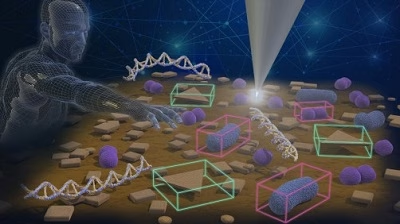
AI-assisted atomic force microscopy identifies tiny surface structures such as nanostructures, DNA assemblies and bacterial cells, and directs follow-up scans to regions most likely to contain scientifically relevant information.
Although atomic force microscopy (AFM) reveals structures as small as molecules, operating the instrument still requires expert judgment about where to scan, how to adjust settings and which features deserve closer study. SimuScan reduces that burden, making AFM faster, more consistent and better suited for high-throughput research.
In a paper published in Nature Communications, the researchers describe how SimuScan addresses one of AI’s biggest obstacles for AFM: the shortage of high-quality labeled training data.
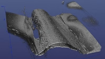
At first glance, interpreting AFM images looks like a standard image-analysis problem. But AFM images differ fundamentally from photographs because they reflect both the sample and the measurement process.
Millan Solsona put it simply: “Tip geometry, drift, flattening and contamination can all introduce artifacts that resemble real nanoscale structures. Experienced users learn to distinguish them; AI models must be taught to do the same.”
SimuScan tackles the data problem by generating synthetic AFM images, along with automatic labels tied directly to the simulated object geometry, so models can be trained without large volumes of hand-annotated experimental data.
To work in real laboratories, however, the synthetic images must be realistic. Rather than producing pristine images, SimuScan recreates the imperfections AFM users encounter every day—including tip effects, scanner drift, electronic noise, contamination and surface roughness.
The researchers validated SimuScan by training AI models on synthetic images and testing whether they could accurately identify features in real AFM data.
With SimuScan, much of that burden shifts to computation. The system can generate large datasets, including thousands of labeled images, with controlled variability in object shapes, backgrounds and artifacts. In this approach, experimental data are used primarily to test and refine the models rather than create most of the training labels.
Beyond analyzing images after the scan, SimuScan supports a closed-loop approach to targeted imaging. The process starts with a fast, low-resolution survey scan across a relatively large area. The AI then identifies and segments features, ranks potential targets based on user-defined criteria and directs the microscope to regions most likely to contain scientifically relevant information. The process can repeat as needed.
In demonstrations spanning fabricated nanostructures, DNA assemblies, and bacterial cells, the team showed that models trained primarily on synthetic data could transfer to real AFM images.
For SimuScan to become commonplace, the researchers said it must integrate more seamlessly with microscope software and include straightforward ways to verify that AI models remain reliable as instruments and experimental conditions change.
The first applications are likely to involve studies requiring measurements of thousands of similar objects, such as nanoparticles, DNA nanostructures and bacterial cells.
In the long run, the researchers envision microscopes becoming active partners in discovery rather than passive imaging tools. SimuScan is one step toward that future.
For more information
Oak Ridge National Laboratory
https://www.ornl.gov/