Imagine a camera so powerful it could take freeze-frame photographs of a moving electron—an object traveling so fast it could circle the Earth many times in a matter of a second. Researchers at the University of Arizona (U of A) have developed the world’s fastest electron microscope that can do just that.
Mohammed Hassan, associate professor of physics and optical sciences, led a team of researchers in his department that published the research article “Attosecond electron microscopy and diffraction” in the Science Advances journal.
In ultrafast electron microscopes, instead of relying on the speed of a camera’s shutter to dictate image quality, the resolution of a transmission electron microscope is determined by the duration of electron pulses. The faster the pulse, the better the image.
Ultrafast electron microscopes previously operated by emitting a train of electron pulses at speeds of a few attoseconds. An attosecond is one quintillionth of a second. Pulses at these speeds create a series of images, like frames in a movie—but scientists were still missing the reactions and changes in an electron that takes place in between those frames as it evolves in real time.
In order to see an electron frozen in place, U of A researchers, for the first time, generated a single attosecond electron pulse, which is as fast as electrons move, thereby enhancing the microscope’s temporal resolution, like a high-speed camera capturing movements that would otherwise be invisible.
U of A researchers developed a microscope in which a powerful laser is split and converted into two parts—a very fast electron pulse and two ultra-short light pulses. The first light pulse, known as the pump pulse, feeds energy into a sample and causes electrons to move or undergo other rapid changes.
The second light pulse, also called the “optical gating pulse” acts like a gate by creating a brief window of time in which the gated, single attosecond electron pulse is generated. The speed of the gating pulse therefore dictates the resolution of the image. By carefully synchronizing the two pulses, researchers control when the electron pulses probe the sample to observe ultrafast processes at the atomic level.
“These movements happen in attoseconds. But now, for the first time, we are able to attain attosecond temporal resolution with our electron transmission microscope—and we coined it ‘attomicroscopy.’ For the first time, we can see pieces of the electron in motion,” Hassan said.
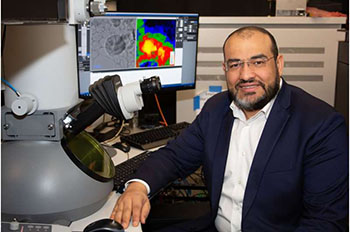
Image – Mohammed Hassan, associate professor of physics and optical sciences at the University of Arizona, led a group of researchers in developing the first transmission electron microscope powerful enough to capture images of electrons in motion. Courtesy of Amee Hennig.
************
For more information:
University of Arizona