Researchers at the Swiss Light Source (SLS) recently developed a pioneering x-ray technique to probe the 3D orientation of a material’s building blocks at the nanoscale. Applied to a polycrystalline catalyst, the technique allows the visualization of crystal grains, grain boundaries and defects—key factors dictating catalyst performance. Beyond catalysis, the innovation unlocks previously inaccessible details about the structure of diverse functional materials in numerous applications.
The grains of these materials are often tiny: tens of nanometers in size. And it is their arrangement in three-dimensions over extended volumes that is property-determining. Yet until now, techniques to probe the organization of materials at the nanoscale have largely been confined to two-dimensions or are destructive in nature.
Now, using x-rays generated by the SLS, a collaborative team of researchers from Paul Scherrer Institute (PSI), ETH Zurich, the University of Oxford and the Max Plank Institute for Chemical Physics of Solids have succeeded in creating an imaging technique to access this information in three-dimensions.
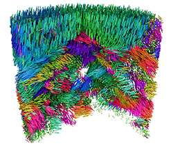
Their technique is known as x-ray linear dichroic orientation tomography, or XL-DOT for short. XL-DOT uses polarized x-rays from the SLS, to probe how materials absorb x-rays differently depending on the orientation of structural domains inside. By changing the polarization of the x-rays, while rotating the sample to capture images from different angles, the technique creates a three-dimensional map revealing the internal organization of the material.
The team applied their method to a chunk of vanadium pentoxide catalyst about one micron in diameter, used in the production of sulfuric acid. Here, they could identify minute details in the catalyst`s structure including crystalline grains, boundaries where grains meet, and changes in the crystal orientation. They also identified topological defects in the catalyst. Such features directly affect the activity and stability of catalysts, so knowledge of this structure is crucial in optimizing performance.
“Linear dichroism has been used to measure anisotropies in materials for many years, but this is the first time it has been extended to 3D. We not only look inside, but with nanoscale resolution,” says Valerio Scagnoli, senior scientist in the Mesoscopic Systems, a joint group between PSI and ETH Zurich. “This means that we now have access to information that was not previously visible, and we can achieve this in small but representative samples, several micrometers in size.”
Although the researchers first had the idea for XL-DOT in 2019, it would take another five years to put it into practice. Together with complex experimental requirements, a major hurdle was extracting the three-dimensional map of crystal orientations from terabytes of raw data. This mathematical puzzle was overcome with the development of a dedicated reconstruction algorithm by Andreas Apseros, first author of the study, during his doctoral studies at PSI, funded by the Swiss National Science Foundation (SNSF).
Given the nondestructive nature of XL-DOT, the researchers foresee operando investigations of systems such as batteries as well as catalysts. “Catalyst bodies and cathode particles in batteries are typically between ten and fifty micrometers in size, so this is a reasonable next step,” says Johannes Ihli, formerly of cSAXS and currently at the University of Oxford, who led the study.
Yet the new technique is not just useful for catalysts, the researchers emphasize. It is useful for all types of materials that exhibit ordered microstructures, whether biological tissues or advanced materials for information technology or energy storage.
Article source – Miriam Arrell, Paul Scherrer Institute.
Image – Many functional materials are composed of coherent grains, where molecules and atoms are arranged in a repeating pattern that determines performance. X-ray linear dichroic orientation tomography (XL-DOT) allows 3D mapping of material microstructure at the nanoscale. Here, the technique is applied to a pillar of vanadium pentoxide catalyst, used in the production of sulfuric acid. The colors in the tomogram represent the different orientation of grains. Courtesy of Andreas Apseros, Paul Scherrer Institute.
************
For more information:
Swiss Light Source SLS