A team of international researchers have discovered a novel photothermal coherence tomography technique—frequency-multiplexed photothermal correlation tomography (FM-PCT)—that brings infrared thermography (IRT) from limited 2D imaging to 3D tomography.
Infrared thermography (IRT) is an effective inspection technique in manufacturing due to its contactless and noninvasive imaging mode. However, existing IRT techniques can only produce 2D results of subsurface structures. The limitations of finite 2D imaging modes significantly impede the inspection and evaluation of material aging and failure.
The research collaboration involving Laval University, Harbin Institute of Technology, University of Toronto, University of L’Aquila, and University of Rome introduced the high-resolution photothermal tomographic technique that can detect subsurface 3D structures of materials with precision comparable to x-ray micro-computed tomography.
“Traditional diffusion-wave techniques are limited by the physics of parabolic diffusion and can only produce depth-integrated planar images,” said Andreas Mandelis, corresponding author on the paper and Professor at the University of Toronto. “We need to design a novel imaging modality that can preserve the energy within the instantaneous frequency bandwidth with minimal or no loss, despite the diffusive nature of the signal.”
Infrared thermography is based on the photothermal effect in materials, i.e., an abnormal thermal distribution when a heat wave encounters a discontinuous interface. Multiple algorithms have been developed to quantitatively estimate defect depths or sizes by assuming regular defect shapes and constant depths, making them less effective for real-world applications.
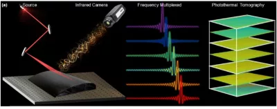
In FM-PCT, a single pulse or line-scan laser is applied to the specimen. According to Fourier transform theory, single pulse excitation can be treated as a combination of multifrequency signals. This provides a new perspective on the truncated-correlation process.
The FM-PCT modality decomposes the pulse excitation into multiple sinusoidal signals and performs matched filtering with captured thermal signals. As is well known, modulation frequency is directly related to the penetration depth. Therefore, by controlling the frequency of matched filtering, FM-PCT can create tomographic images at various subsurface depths.
“FM-PCT bridges the gap in microscale tomography caused by the limitations of general x-ray CT and ultrasound imaging, especially for thin specimens,” said co-author Hai Zhang, Full Professor at Harbin Institute of Technology and Adjunct Professor at Laval University. “This technique will significantly improve the inspection capability and help detect early-stage defects in industrial manufacturing and lesions in biomedical fields.”
The team’s work was published in the International Journal of Extreme Manufacturing.
Image – By employing either flash pulse excitation or linear laser scanning, thermophotonic energy penetrates the material’s interior and evolves with time. The acquired relaxation signals are then processed through truncated correlation using frequency-incremented units. Courtesy of the International Journal of Extreme Manufacturing, 2025, doi.org/10.1088/2631-7990/ada837.
************
For more information:
International Journal of Extreme Manufacturing