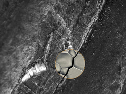
A Texas A&M University research team has taken the first 3D image of a microscopic crack propagating through a metal damaged by hydrogen. Previously, the only way to analyze this kind of metal failure was to look at the separated pieces of a completely fractured component, and add some educated guesses to the failure analysis.
The new research shows what is happening at the crack tip as a part begins to fracture. The team identified 10 microscopic structures that make metals stronger and less susceptible to hydrogen embrittlement. Scientists have studied this process for over 150 years, but it remains difficult to predict because the mechanisms behind it are not yet completely understood.
Researchers employed two different synchrotron tools—high-energy diffraction microscopy and x-ray absorption tomography—to analyze the microscopic structure of a crack in a nickel-base superalloy. The team was able to show not only which grain boundaries are stronger but also the specific properties that improve their performance. This finding could ultimately allow engineers to build stronger metals by designing them with those characteristics.
************
Image – Advanced imaging provides clues to predicting fracture in metals. Courtesy of Texas A&M.
For more information: