Researchers at the University of Michigan have developed a technique that allows the study of microstructures inside metals, ceramics, and rocks using X-rays in a standard laboratory, eliminating the need to travel to a particle accelerator. This advancement makes 3D X-ray diffraction (3DXRD) more accessible, enabling rapid analysis of samples and prototypes in both academic and industrial settings, and providing more opportunities for student involvement. 3DXRD works by reconstructing 3D images from X-rays taken at multiple angles, similar to a CT scan, but with the material sample rotating in front of a powerful beam that emits about a million times more X-rays than a medical X-ray.
The huge X-ray concentration produces a micro-scale image of the tiny fused crystals that make up most metals, ceramics and rocks, known as polycrystalline materials.
Results help researchers understand how materials react to mechanical stresses by measuring thousands of individual crystals’ volume, position, orientation and strain. For example, imaging a sample from a steel beam under compression can show how crystals respond to bearing the weight of a building, helping researchers understand large-scale wear.
Synchrotrons were once the only facilities able to produce enough X-rays for 3DXRD as electrons spit off scads of X-rays as they travel through circular particle accelerators, which can then be directed into a sample.
While synchrotron X-ray beams produce state-of-the-art detail, there are only about 70 facilities worldwide. Research teams must put together project proposals for “beam time.” Accepted projects often must wait six months to up to two years to run their experiments, which are limited to a maximum of six days.
In an effort to make this technique more widely available, the research team worked with PROTO Manufacturing to custom build the first laboratory-scale 3DXRD. As a whole, the instrument is about the size of a residential bathroom, but could be scaled down to the size of a broom closet.
“This technique gives us such interesting data that I wanted to create the opportunity to try new things that are high risk, high reward and allow teachable moments for students without the wait-time and pressure of synchrotron beam time,” said Ashley Bucsek, U-M assistant professor of mechanical engineering and materials science and engineering and co-corresponding author of the study published in Nature Communications.
Previously, small-scale devices could not produce enough X-rays for 3DXRD because at a certain point, the electron beam pumps so much power into the anode—the solid metal surface that the electrons strike to make X-rays—that it would melt. Lab-3DXRD leverages a liquid-metal-jet anode that is already liquid at room temperature, allowing it to take in more power and produce more X-rays than once possible at this scale.
The researchers put the design to the test by scanning the same titanium alloy sample using three methods: lab-3DXRD, synchrotron-3DXRD and laboratory diffraction contrast tomography or LabDCT—a technique used to map out crystal structures in 3D without strain information.
Lab-3DXRD was highly accurate, with 96% of the crystals it picked up overlapping with the other two methods. It did particularly well with larger crystals over 60 micrometers, but missed some of the smaller crystals. The researchers note that adding a more sensitive photon-counting detector, which detects the X-rays that are used to build the images, could help catch the finest-grained crystals.
With this technique available in-house, Bucsek’s research team can try new experiments, honing parameters to prepare for a larger experiment at a synchrotron.
“Lab-3DXRD is like a nice backyard telescope while synchrotron-3DXRD is the Hubble Telescope. There are still certain situations where you need the Hubble, but we are now well prepared for those big experiments because we can try everything out beforehand,” Bucsek said.
Beyond enabling more accessible experiments, lab-3DXRD allows researchers to extend projects past the synchrotron six day limit, which is particularly helpful when studying cyclic loading—how a material responds to repeated stresses over thousands of cycles.
For more information: The Michigan Center for Materials Characterization
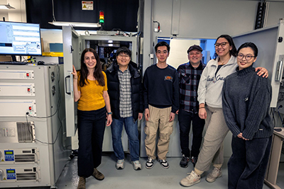
Image: Members of Professor Ashley Bucsek’s lab group are able to use three-dimensional X-ray diffraction to study polycrystalline materials on campus, a technique previously only available in specialized synchrotron facilities. Left to right: Ashley Bucsek, Sangwon Lee, Wenxi Li, Abdulhamit Sarac, Janice Moya and Yuefeng Jin. Image credit: Marcin Szczepanski, Michigan Engineering