In a feat requiring perseverance, world-leading technology, and extreme caution, scientists have used intense x-rays to inspect irradiated nuclear fuel. The imaging, led by researchers at Purdue University and conducted at the U.S. Department of Energy’s (DOE) Argonne National Laboratory, revealed a 3D view of the fuel’s interior structure, laying the groundwork for better nuclear fuel designs and models.
Until now, examinations of uranium fuel have been limited to mostly surface microscopy or to various characterization techniques using mock versions that possess little radioactivity. But scientists want to know at a deeper level how the material changes as it undergoes fission inside a nuclear reactor. The resulting insights from this study can lead to nuclear fuels that function more efficiently and cost less to develop.
To get an interior view of the uranium-zirconium fuel studied, the researchers sectioned off a bit of used fuel small enough to be handled safely. Then, to see inside this tiny metallic sample, they turned to the Advanced Photon Source (APS), a DOE Office of Science User Facility located at Argonne.
Before the researchers could approach the formidable task of isolating a fuel sample and placing it under an x-ray beam, they needed to find the right specimen. Exploring fuels archived at DOE’s Idaho National Laboratory, they identified a uranium-zirconium fuel that spent a total of two years at full power in the Fast Flux Test Facility in Hanford, Washington, and was removed from the reactor in the early 1990s. They had to wait decades for the fuel to radiologically cool or decay.
To reduce the radioactivity, the researchers used a focused ion beam with scanning electron microscopy to create a much smaller sample. The tool allowed them to pinpoint an area of interest and deploy a stream of ions that essentially milled out a cube of material. The resulting sample was roughly 100 microns across, no larger than the diameter of a human hair.
The Purdue research team then worked with Argonne scientists at beamline 1-ID-E, a high-brilliance X-ray source at the APS, to examine the sample. Their technique used micro computed tomography (CT), which detects at high resolution the x-ray beam as it emerges on the other side of the sample. From multiple images taken as the fuel was rotated, computers could reconstruct its internal features based on how it altered the incoming beam, similar to a medical CT scan.
In particular, the researchers were interested in the phenomenon of swelling. The safety and longevity of any given nuclear fuel depends on being able to predict how much it will swell. Too much swelling can cause the uranium to react with, and possibly fracture, its protective outer layer, called a cladding.
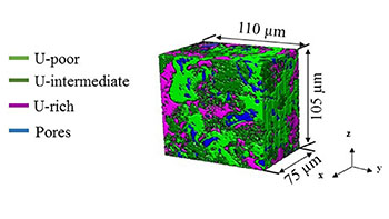
In addition to providing a clearer, localized picture of the fuel structure and the different material phases that developed over time, the study at the APS revealed evidence that the release of fission gases might continue to occur beyond the thresholds assumed in previous analyses. This type of data can help strengthen fuel performance codes, which in turn would help lower the cost of fuel development, since reliable computer simulations can minimize the number of expensive irradiation tests needed.
Image – 3D image reconstruction of a sample of irradiated nuclear fuel, showing the three thresholded uranium phases co-existing with pores. Courtesy of Maria Okuniewski/Purdue University.
************
For more information: