Researchers at the Max Planck ETH Centre for Learning Systems, Germany, have developed an imaging technique that for the first time recognizes cell-sized microrobots individually and at high resolution in a living organism.
With the potential to revolutionize medicine, removing blood clots from the brain without any major surgical intervention or delivering drugs precisely into a diseased organ that is difficult to reach, tiny medical microrobotics promise to fundamentally change future medical treatments.
Researchers believe the circulatory system might serve as an ideal delivery route for the microrobots since it reaches all organs and tissues in the body, so the microrobots must not be larger the smallest blood vessels. In humans, the average capillary diameter is only 8 micrometers.
Such a small size makes the microrobots invisible to the naked eye, and science has not found a technical solution to detect and track the micron-sized robots individually as they circulate in the body, until now. Tracking is absolutely necessary, and without imaging, microrobotics is essentially blind. Real-time, high-resolution imaging is essential for detecting and controlling cell-sized microrobots in a living organism.
Imaging is also a prerequisite for monitoring therapeutic interventions performed by the robots and verifying that they have carried out their task as intended. According to researchers, the lack of ability to provide real-time feedback on microrobots has been a major obstacle on the way to clinical application.
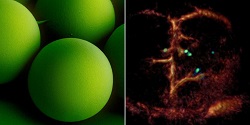
The research team developed a non-invasive imaging technique that for the first time was able to clearly detect and track tiny robots as small as five micrometers in real time in the brain vessels of mice.
A dedicated optoacoustic tomography technology was used to detect the tiny robots one by one, in high resolution and in real time. This unique imaging method makes it possible to detect the tiny robots in deep and hard-to-reach regions of the body and brain, which would not have been possible with optical microscopy or any other imaging technique.
In optoacoustics, light is first emitted and absorbed by the respective tissue. The absorption then produces tiny ultrasound waves that can be detected and analyzed to result in high-resolution volumetric images.
To make the microrobots highly visible in the images, the researchers needed a suitable contrast material. For their study, they used spherical, silica particle-based microrobots with a Janus-type coating. This type of robot has a very robust design and is very well qualified for complex medical tasks. It is named after the Roman god Janus, who had two faces. In the robots, the two halves of the sphere are coated differently.
In this study, the researchers coated one half of the robot with nickel and the other half with gold to be detected individually with the new optoacoustic imaging technique. The gold provides suitable contrast and also allows minimizing the cytotoxic effect of the nickel coating. If in the future microrobots are to operate in living animals or humans, they must be made biocompatible and non-toxic, which is part of an ongoing research.
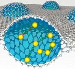
In addition to gold, the researchers also tested the use of small bubbles called nanoliposomes, which contained a fluorescent green dye that also served as a contrast agent. Liposomes also have the advantage that they can be loaded with potent drugs, important for future approaches to targeted drug delivery.
Nickel was used as a magnetic drive medium and a simple permanent magnet to pull the robots. In follow-up studies, the group wants to test the optoacoustic imaging with more complex manipulations using rotating magnetic fields, to provide the ability to precisely control and move the microrobots even in strongly flowing blood.
Image – A breakthrough: tiny circulating microrobots, which are as small as red blood cells (left), were visualized one-by-one in the blood vessels of mice with optoacoustic imaging (right). Courtesy of: ETH Zurich / Max Planck Institute for Intelligent Systems.
For more information:
Max Planck ETH Centre for Learning Systems