Researchers from the University of Warsaw, in cooperation the Weizmann Institute of Science in Israel, have introduced a new technique of super-resolution microscopy, called super-resolution optical fluctuation image scanning microscopy (SOFISM). Their new method of microscopy, in theory, has no resolution limit. The team has demonstrated a fourfold improvement over the diffraction limit in practice.
In SOFISM, the naturally occurring fluctuations in emission intensity of fluorescent markers are used to further enhance the spatial resolution of an image scanning microscope (ISM). ISM, an emerging super-resolution method, has already been implemented in commercial products and proven valuable especially for the bio-imaging community. Largely, since it achieves a modest improvement in lateral resolution (x2), with very few changes to the optical setup and without the common handicap of long exposure times. Thus, it enables a natural extension of the capabilities of a standard confocal microscope. ISM uses a confocal microscope in which a single detector is replaced with a detector array.
In SOFISM correlations of intensities detected by multiple detectors are computed. In principle, the measurement of the n-th order correlation can lead to a factor of 2n resolution improvement with respect to the diffraction limit. In practice, the resolution achievable for higher-order correlations is limited by the signal-to-noise ratio of the measurements.
“SOFISM is a compromise between ease of use and resolution. We believe that our method will fill the niche between the complex, difficult-to-use techniques providing very high resolution and the easy-to-use lower-resolution methods. SOFISM does not have a theoretical resolution limit, and in our article, we demonstrate results which are four times better than the diffraction limit. We also show that the SOFISM method has a high potential in the imaging of three-dimensional biological structures,” said Dr. Radek Lapkiewicz from the Quantum Optics Lab at the University of Warsaw.
Crucially, SOFISM is highly accessible, as it only requires introducing a small modification to the widely-used confocal microscope—replacing its photomultiplier tube with a SPAD array detector. In addition, it is necessary to slightly increase the measurement time and change the data processing procedure. “Until recently, SPAD array detectors were expensive and their specifications were not sufficient for correlation-based microscopy. This situation has recently changed. The new SPAD detectors introduced last year removed both the technological and price-related barriers. This makes us think that fluorescence microscopy techniques such as SOFISM might, in a few years’ time, become widely used in the field of microscopic examination,” stressed Dr. Lapkiewicz.
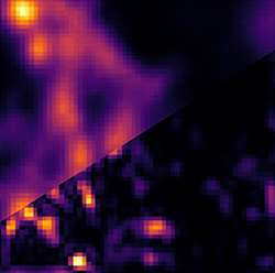
Image – Image of microtubules in a fixed cell sample. A 3 μm x 3 μm confocal scan of microtubules in a fixed 3T3 cell labelled with quantum dots analyzed in two ways. Upper left: image scanning microscopy (ISM), lower right: super-resolution optical fluctuation image scanning microscopy (SOFISM) after Fourier-reweighting. Courtesy of UW Physics/A. Makowski.
************
For more information: