The Korea Advanced Institute of Science and Technology (KAIST) announced that a joint research team led by Professor YongKeun Park of the Department of Physics and Dr. Jun Lim of the Pohang Accelerator Laboratory has succeeded in developing a core technology that can overcome the resolution limitations of existing x-ray microscopes.
This study, in which Dr. KyeoReh Lee participated as the first author, was published in Light: Science and Application (“Direct High-resolution X-ray Imaging Exploiting Pseudorandomness”).
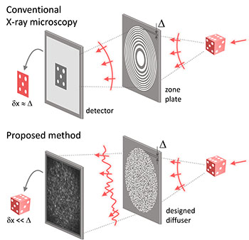
X-ray nanomicroscopes do not have refractive lenses. In an x-ray microscope, a circular grating called a concentric zone plate is used instead of a lens. The resolution of an image obtained using the zone plate is determined by the quality of the nanostructure that comprises the plate. There are several difficulties in fabricating and maintaining these nanostructures, which set the limit to the level of resolution for x-ray microscopy.
The research team developed a new x-ray nanomicroscopy technology to overcome this problem. The x-ray lens proposed by the research team is in the form of numerous holes punched in a thin tungsten film, and generates random diffraction patterns by diffracting incident x-rays. The research team mathematically identified that, paradoxically, the high-resolution information of the sample was fully contained in these random diffraction patterns, and actually succeeded in extracting the information and imaging the internal states of the samples.
The first author and one of the co-corresponding author, Dr. KyeoReh Lee of KAIST Department of Physics, said, “In this study, the resolution was limited to 14 nm, but if the next-generation x-ray light source and high-performance x-ray detector are used, the resolution would exceed that of the conventional x-ray nano-imaging and approach the resolution of an electron microscope.” and added, “Unlike an electron microscope, x-rays can observe the internal structure without damaging the sample, so it will be able to present a new standard for non-invasive nanostructure observation processes such as quality inspections for seimconductors.”
Image – Designed diffuser as x-ray imaging lens. (a) Schematic of full-field transmission x-ray microscopy. The attenuation (amplitude) map of a sample is measured. The image resolution (dx) is limited by the outermost zone width of the zone plate (D). (b) Schematic of the proposed method. A designed diffuser is used instead of a zone plate. The image resolution is finer than the hole size of the diffuser (dx << D). Courtesy of KAIST.
************
For more information:
KAIST (Korea Advanced Institute of Science and Technology)