Speckle-MAIN technology, developed by researchers at the University of California, San Diego, involves a specially engineered material that shortens the wavelength of light as it illuminates the sample.
Conventional light microscopes have a resolution limit of 200 nanometers (nm), meaning that any objects closer than this distance will not be observed as separate objects. While there are more powerful tools out there such as electron microscopes, which have the resolution to see subcellular structures, they cannot be used to image living cells because the samples need to be placed inside a vacuum chamber.
“The major challenge is finding one technology that has very high resolution and is also safe for live cells,” said Professor Zhaowei Liu, a researcher in the Department of Electrical and Computer Engineering, Material Science and Engineering Program, and the Center for Memory and Recording Research at the University of California, San Diego.
With the speckle-MAIN technology, a conventional light microscope can be used to image live subcellular structures with a resolution of up to 40 nm. The technology consists of a microscope slide that’s coated with a type of light-shrinking material called a hyperbolic metamaterial. It is made up of nanometers-thin alternating layers of silver and silica glass. As light passes through, its wavelengths shorten and scatter to generate a series of random high-resolution speckled patterns.
When a sample is mounted on the slide, it gets illuminated in different ways by this series of speckled light patterns. This creates a series of low-resolution images, which are all captured and then pieced together by a reconstruction algorithm to produce a high-resolution image.
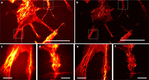
Professor Liu and colleagues tested their technology with a commercial inverted microscope. They were able to image fine features, such as actin filaments, in fluorescently labeled Cos-7 cells — features that are not clearly discernible using just the microscope itself. The technology also enabled the scientists to clearly distinguish tiny fluorescent beads and quantum dots that were spaced 40 to 80 nm apart.
The team’s work was published in the journal Nature Communications.
For more information: Nature Communications Journal