Sandia National Laboratories, Albuquerque, N.M., has borrowed and refined X-ray phase contrast imaging, originally developed for medical X-rays, to look inside low-density materials without taking them apart. These materials include laminate layers in airplane wings, to foams and epoxies that cushion parts.
X-ray phase contrast imaging measures not only the number of X-ray photons that get through the sample, as in conventional X-ray imaging, but also the phase of the X-rays after they pass through, offering a complete look at interfaces inside a structure.
“For low-density materials such as plastics, polymers, foams, and other encapsulants, this phase signal can be a thousand times bigger than the absorption signal (of conventional X-rays),” says principal investigator Amber Dagel.
X-ray phase contrast imaging could inspect microfabrication packaging, integrated circuits, or micro-electro-mechanical components, and could be used to study ceramics, polymers, chemicals or explosives.
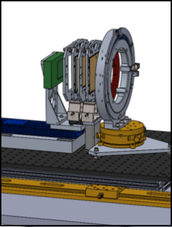
Sandia’s technique achieved X-ray phase contrast imaging in a lab without a synchrotron, an expensive piece of equipment the size of a football field. Gratings, optical components that look like bunches of upright parallel bars, create interference in the X-ray beam, like an interferometer, merging sources of light to create an interference pattern that can be measured.
Gratings are critical to the technique, and using them at higher energies “will let us look at more samples, samples that are denser or samples that are bigger,”Dr. Dagel said. They’re difficult to make, but Sandia’s metal micromachining team led by Christian Arrington makes highly uniform gratings up to four inches square. That’s considered large scale, and Sandia is able to make gratings as one large piece with good uniformity. Grating size determines how much of a sample can be seen at once.
Photo shows the sample that sits in a ring. Behind the sample are phase and analyzer gratings and the detector.