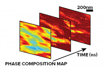
In a new study, researchers from Argonne National Laboratory have for the first time been able to look at a structural phase transition in minute detail on a very fast timescale. The scientists made a series of x-ray images that are spaced less than one-tenth of 1 billionth of a second apart using nanodiffraction microscopy. Scientists plan to use phase transitions to be able to control the electronic, structural or magnetic properties of different materials as they undergo these changes, such as for use in new types of computer memories.
“A typical video might play at 30 frames per second, so this is approximately a slow-motion video that can resolve dynamics that are extremely fast,” said Haidan Wen, a physicist at the U.S. Department of Energy’s (DOE) Argonne National Laboratory.
The ability to witness the evolution of material behavior with such precision in time and space has revealed unusual behaviors in certain materials that undergo a phase change, including many magnetic materials.
We’re able to zoom into a sample in terms of time and space in ways we have never been able to before,” said Youngjun Ahn, the first author of the study.
Ahn is a former graduate student intern at Argonne from the University of Wisconsin-Madison. For this work, he collaborated closely with Wen. “This method gives us a precise view of structural changes in our sample that are challenging to see with any other method,” Ahn said.
The study used the Hard X-Ray Nanoprobe operated by the Center for Nanoscale Materials (CNM) at the Advanced Photon Source (APS) at Argonne. The APS and CNM are DOE Office of Science user facilities.
In looking at phase transitions in an iron-rhodium compound, the researchers found a way to watch the structure of the compound change between two magnetic configurations. The change causes an expansion of the atomic network that is very small—but enough to have significant consequences for the magnetism.
“One of the things that’s very interesting about this particular material—iron-rhodium—is that it has a phase transition at a temperature that could be used for these kinds of applications,” said University of Wisconsin-Madison professor Paul Evans. “But in order to do the kinds of manipulations we’re interested in, we need a better ‘camera.’ That’s why using this newly developed technique to study it is important.”
The upcoming upgrade to the APS will have significant implications for further experiments visualizing these kinds of phase transitions. “After the APS upgrade,” said Argonne x-ray scientist Martin Holt, “we expect to achieve higher spatial resolution, in particular, by exploiting the enhanced coherence of the x-ray beam. Our development of ultrafast time resolution within that type of x-ray microscopy is what helps us understand the causes of the types of effects we’re observing. This is a unique capability that the upgraded APS can offer.
Their work was published in PNAS, “X-ray nanodiffraction imaging reveals distinct nanoscopic dynamics of an ultrafast phase transition.”
Image – X-ray nanodiffraction maps of the ferromagnetic phase transition. Courtesy of Haidan Wen, Argonne National Laboratory.
************
For more information: