Andrew Woodard
Semiconductor Insights, Ontario, Canada
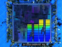
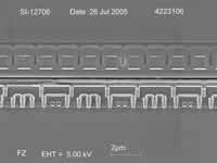
Light diffracting off decapped microcontroller
2nd Place:
Wong Yaw Yuan
Micron Semiconductor, Asia
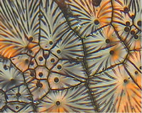
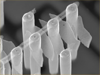
Leaf pattern with bubble like void observed on
the die attached tape after delamination occur
due to insufficient cure of polyimide.
3rd Place:
Mark Lam Tiam Weng
Institute of Microelectronics A-Star, Singapore
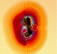
Electronic Foetus – Birth of a chip. Defect on ECP copper before CMP looks like a foetus developing.
Category II: Black and White Images
1st Place:
Gil Garteiz
Tower Semiconductor, Israel
A 22 angstrom low-voltage gate ruptured by ESD testing is exposed from the backside by removing the substrate. The final wet etch step has etched the gate poly out from behind the rupture oxide. Layers shown: STI, silicided active & contacts, spacers, gate oxide
2nd Place:
Frank Zhenge
Semiconductor Insights, Ontario, Canada
SRAM cross section
3rd Place:
Sue Li Poh
Micron Semiconductor, Asia
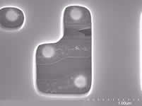
Contamination at Contact Plugs
Category III: False Color Images
1st Place:
Robert Ulfig
Imago Scientific Instruments, Wisconsin, USA
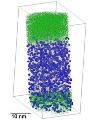
3D Atom Probe Analysis of CVD Deposited Silicon Germanium, Boron Doped Silicon and Boron Doped Silicon Germanium films. Each dot or sphere indicated the location of individual atoms in the films. Silicon(Grey), Germanium(Green), and Boron(Blue Spheres). Only a fraction of the atoms are displayed for clarity.
2nd Place:
Kartik Ramanujachar
Texas Instruments, Texas, USA
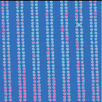
Scanning current image of a SRAM showing
gate contact leakage that caused a single bit failure.
The leaky gate contact appears like an extra
contact juxtaposed between four other contacts.
3rd Place:
Bernhard Tittmann
Pennsylvania State University, USA
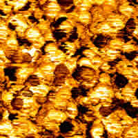
Image obtained with Ultrasonic Atomic Force Microscope which images relative modulus changes. The sample is a “green body” of Alumina particles (orange-white) with 3% binder (dark circles) The binder (Acrylic Emulsion) shows up as dark, because it has a lower modulus than the Alumina. The image is 10×10 micron. The particle size is about 1 micron. The binder could not be distinguished from the Alumina by any other microscope available.